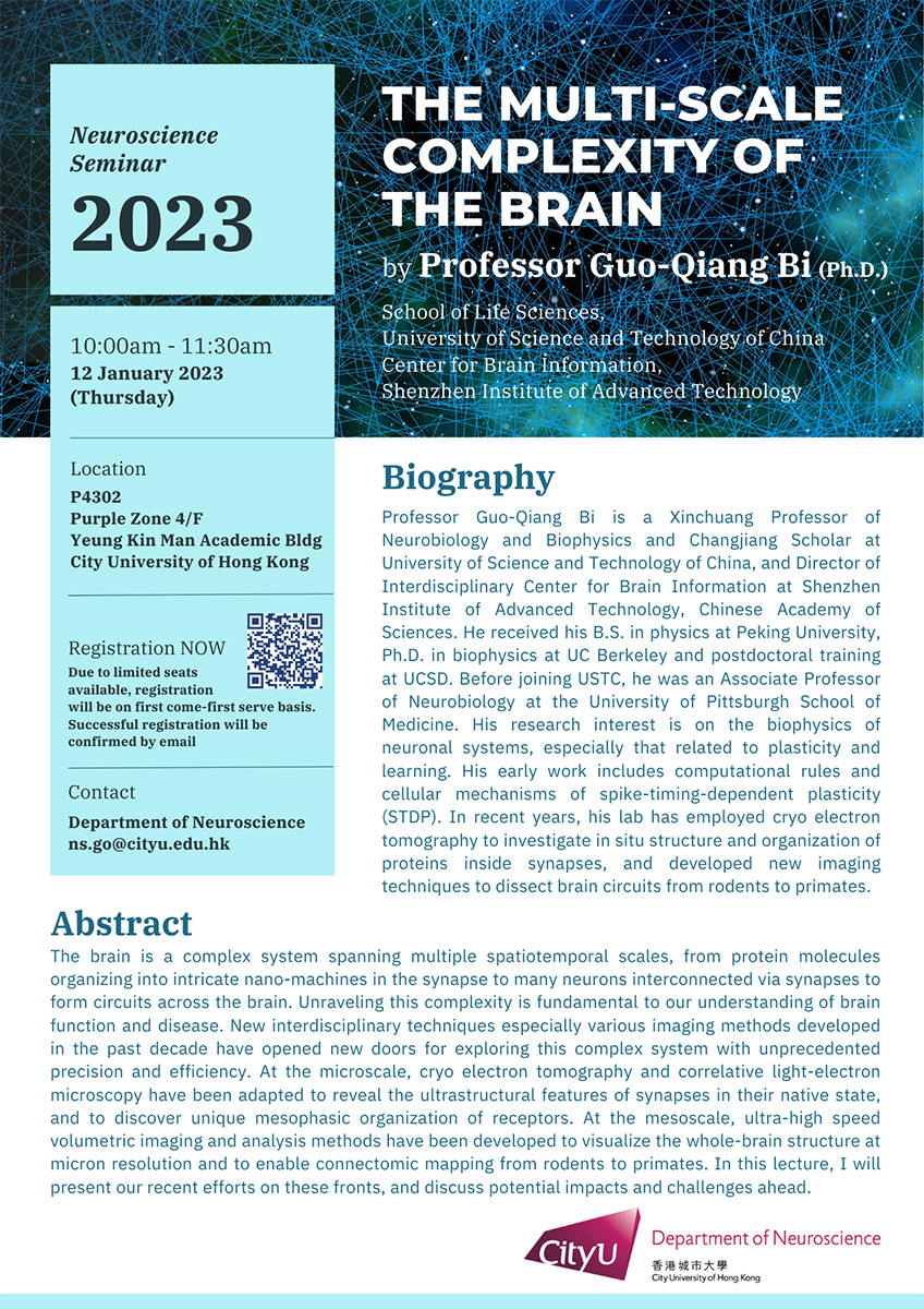
On 12th January, the Department of Neuroscience organised a seminar and invited Professor Guo-Qiang Bi, Xinchuang Professor of Neurobiology and Biophysics and Changjiang Scholar at the University of Science and Technology of China and director of the Interdisciplinary Center for Brain Information at Shenzhen Institute of Advanced Technology, Chinese Academy of Sciences, to deliver a seminar titled “The Multi-scale Complexity of the Brain”.
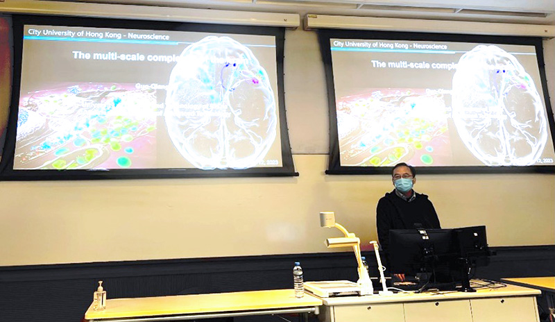
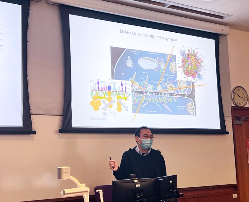
He reviewed their recent efforts on imaging methods for exploring the brain’s connectome from rodents to primates in multiscale. At the microscale, cryoelectron tomography and Correlative Light-Electron Microscopy have been adapted to reveal the ultrastructural features of synapses in their native state and to discover the unique mesophasic organisation of receptors. At the mesoscale, ultra-high-speed volumetric imaging and analysis methods have been developed to visualise the whole-brain structure at micron resolution and to enable connectomic mapping from rodents to primates.
Professor Bi presented an approach that combines primate-optimised tissue sectioning and clearing with ultrahigh-speed fluorescence microscopy implementing improved volumetric imaging with synchronised on-the-fly-scan and readout (VISoR) technique. They can complete whole-brain imaging of a rhesus monkey at 1 × 1 × 2.5 µm3 voxel resolution within 100 hours. His lab further improved the VISoR system and established a primate-optimised pipeline, including serial sectioning and clearing, 3D Microscopy with semi-Automated Reconstruction and Tracing (SMART). His lab also developed a highly efficient method for long-range tracing of sparse axonal fibres in datasets numbering hundreds of terabytes.
The primate-optimised pipeline may help neuroscientists unveil the mystery of the brain in future works.