Metabolism in Health and Disease
Featured Speaker

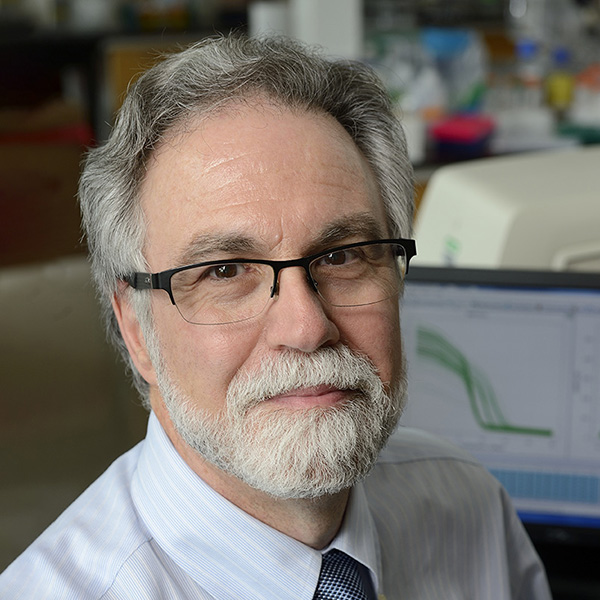
Cancer cells are characterized by high metabolic demand. Oxygen serves as a key substrate in cellular metabolism and bioenergetics. Hypoxia or low oxygen abundance is a common feature of the tumor microenvironment that occurs due to an imbalance between O2 supply and demand. Many of the metabolic responses to hypoxia, which affect levels of glucose, glutamine, glycogen, and lipids, are orchestrated by hypoxia-inducible factors (HIFs), which are O2-regulated transcription factors composed of an O2-labile HIF-α subunit and a constitutively expressed HIF-1β subunit. Increased expression of HIF-1α protein or increased expression of HIF target genes in primary tumor biopsies is associated with increased patient mortality in many types of cancer. HIFs activate the transcription of genes involved in angiogenesis, cancer stem cell specification, immune evasion, invasion and metastasis as well as metabolism. The mechanisms and consequences of homeostatic responses mediated by the HIFs that modulate tumor metabolism will be discussed.
Keynote Speakers

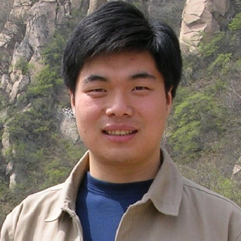
Sufficient energy storage in the form of neutral lipid TAG is important for survival during evolution. However, excess lipid storage leads to the development of metabolic diseases including obesity, diabetes and fatty liver disease. Lipid droplets (LDs) are dynamic subcellular organelles responsible for lipid storage and control intracellular lipid homeostasis. This seminar will discuss the role of CIDE family in controlling LD fusion and lipid storage. CIDE proteins consist Cidea, Cideb and Cidec (Fsp27) are LD and ER-associated proteins. CIDEs deficient animals indicate that these proteins play important roles in controlling lipid storage in adipocytes, hepatocytes, mammary epithelial cells and skin sebocytes. Further molecular and cell biological evidence suggest that CIDE family proteins are highly enriched at LD-LD contact sites (LDCS) and promote atypical form of LD fusion and growth by initiating a directional lipid transfer from smaller to larger LDs. Several regulatory proteins including Perilipin1 (Plin1) and Rab8a are shown to enhance CIDE-mediated LD fusion and growth. Our recent analyses demonstrate that the condensation of Cidec is formed at the LDCS through membrane constrained gel-like phase separation. Using 3D EM tomography and super-resolution imaging, we reveal that Cidec condensates form a gel-like dynamic porous fusion plate with high plasticity contingent on the sizes of the contacting LDs. Thus, we have uncovered the structural and functional significance of phase separation in mediating membrane contact as exemplified by LD fusion and regulating lipid homeostasis. The molecular and physiological insight of lipid droplet fusion and lipid storage will be discovered.

Till June 2022, the pandemic of Coronavirus Disease 2019 (COVID-19) has caused over 532 million infections and over 6.3 million deaths worldwide. It has become the most devastating challenge to global health for a century. As the causative agent of COVID-19, SARS-CoV-2 encodes 16 non-structural proteins (nsp1-nsp16) that assemble a set of protein machineries, the Replication-Transcription Complexes (RTCs), that play central roles in virus replication and transcription cycle inside the host cells.
In the early of COVID-19 outbreak, we rapidly initiated the structural study of SARS-CoV-2 RTCs, aiming to dissect the key mechanisms for SARS-CoV-2 lives in human cells and provide structural information to discover potent antivirals. With great efforts from joint collaborations, we successfully determined the structure of the central RTC (C-RTC) composed by nsp12 (RNA-dependent RNA polymerase, RdRp) with cofactors nsp7 and nsp8, providing the first picture for the world to visualize this key antiviral target. We also elucidated how C-RTC catalyzes and how Remdesivir (RDV) inhibits the synthesis of RNA, through determining the structure of C-RTC in complex with template-product duplex RNA and the active form of RDV. Subsequently, we presented the structure of the elongation RTC (E-RTC), showing how nsp13 (helicase) unwinds the high-ordered structure in genome to yield the functional template for RNA synthesis in C-RTC. After that, we discovered a key intermediate state of RTC towards mRNA capping [Cap(-1)’-RTC], demonstrating the nsp12 NiRAN is indeed the key enzyme to catalyze the second capping action and presenting nsp9 is an “adaptor” for the further recruitments of capping enzymes into RTC. Very recently, we successfully assembled Cap(0)-RTC by Cap(-1)’-RTC and nsp10/nsp14 complex and determined its structures in a monomeric and a dimeric form. The monomeric Cap(0)-RTC structure shows the assembly of a co-transcriptional capping complex (CCC) to RTC for mRNA capping, while most interestingly, the dimeric form reasons an in trans backtracking mechanism for proofreading. Other RTCs responsible for key steps for SARS-CoV-2 living inside cells have also been determined. These works not only provide a basis to understand SARS-CoV-2 proliferates in the host cells through a structural biology lens, but also shed the light for antiviral development against the rapid emerging of SARS-CoV-2 variants.

Cholesterol is an essential lipid and it costs lots of nutrients and energy to make such a molecule. Therefore, mammals increase cholesterol biosynthesis only after feeding and inhibit the process under fasting condition. However, the regulatory mechanisms of cholesterol biosynthesis at fasting-feeding transition are not fully understood. Here we show that the deubiquitylase USP20 stabilizes HMG-CoA reductase (HMGCR), the rate-limiting enzyme in cholesterol biosynthetic pathway, at feeding state. The post-prandially increased insulin and glucose stimulate mTORC1 to phosphorylate USP20 at S132 and S134, which is further recruited to the HMGCR complex and antagonizes its degradation. The feeding-induced stabilization of HMGCR is abolished in the liver-specific Usp20 deficient mice and the Usp20-S132A/S134A knock-in mice. Genetic deletion or pharmacological inhibition of USP20 dramatically decreases diet-induced body weight gain, reduces lipid levels in the serum and liver, improves insulin sensitivity as well as increases energy expenditure. These metabolic improvements by USP20 inhibition are reversed by the constitutively stable HMGCR(K248R). This study reveals an unexpected regulatory axis from mTORC1 to HMGCR through USP20 phosphorylation and demonstrates USP20 inhibitor as a potential cholesterol-lowering drug to treat metabolic diseases including hyperlipidemia, liver steatosis, obesity and diabetes. I will also present our latest findings on cholesterol excretion.

The therapeutic options for diabetic patients with cardiovascular complications are limited, highlighting an outstanding unmet medical need. MG53 (also named TRIM72) is a myokine with cell protective effects. However, MG53 also promotes insulin resistance and metabolic disorders via its E3 ligase activity. Here, we show that in diabetic mice, recombinant E3-dead MG53 mutants (C14A or S255A) effectively protects the heart from ischemia/reperfusion (I/R) injury without metabolic side-effects, whereas wild-type MG53 profoundly exacerbates hyperglycemia and I/R-induced myocardial injury and mortality, especially in mice with advanced diabetes. Consistently, MG53 C14A knock-in mice are protected against high fat diet (HFD)-induced metabolic disorders, while IPC can still trigger cardioprotection in the C14A knock-in mice. These in vitro and in vivo data indicate that E3-dead MG53 mutants not only preserves myocardial protective functions in diabetic individuals, but also defends mice against metabolic dysfunctions incurred by HFD, demonstrating their therapeutic potential in treating diabetes-associated cardiovascular complications.

Voltage-gated sodium (Nav) channels are responsible for the initiation and propagation of action potentials. Being associated with a variety of disorders, Nav channels are targeted by multiple pharmaceutical drugs and natural toxins. We determined the crystal structure of a bacterial Nav channel NavRh in a potentially inactivated state more than a decade ago. Employing the modern methods of cryo-EM, we determined the near atomic resolution structures of a Nav channel from American cockroach (designated NavPaS) and from electric eel (designated EeNav1.4). Most recently, we have determined the cryo-EM structures of representative human Nav channels (Nav1.1/1.2/1.4/1.5/1.7) in complex with distinct auxiliary subunits, toxins, and drugs.These structures reveal the folding principle and structural details of the single-chain eukaryotic Nav channels that are distinct from homotetrameric voltage-gated ion channels. The structures were captured in drastically different states. Whereas the structure of NavPaS has a closed pore and the four VSDs in distinct conformations, the others are semi-open at the intracelluar gate with VSDs exhibiting similar “up”states. The most striking conformational differenc occurs to the III-IV linker, which is essential for fast inactivation. Based on the structural features, we suggest a “door-wedge” allosteric blocking mechanism for fast inactivation of Nav channels. Structural comparison of the conformationally distinct Nav channels provides important insights into the electromechanical coupling mechanism of Nav channels and offers the 3D template to map hundredes of disease mutations.
Invited Speakers

The study of pluripotent stem cells has been mostly focused on molecular biology. However, organelle remodeling and metabolic controlling in cell fate determination remains unclear. After establishing his team since 2010, Prof. Xingguo Liu has been focusing on this direction, and achieved novel findings both in physiological and pathological conditions. On one hand, he discovered the rules of mitochondrial/metabolic regulation of nuclear epigenetics such as the law of mitochondrial oxygen ion signals regulating DNA and histone methylation, a new concept of the " epigenome-metabolome-epigenome" cascade, and organelle remodeling and regulating new functions of pluripotency. On the other hand, he clarified the new pathology of mitochondrial/metabolic diseases such as apoptosis or ferroptosis of liver cells from patients with mitochondrial DNA depletion syndrome, and iPSC technology to drug toxicity research.

Accumulating evidence has demonstrated that immune cells such as macrophages play an important role in regulating the progression of cardiovascular disease and repair. After injury, danger signals released by the damaged tissues trigger the initial pro-inflammatory phase essential for removing cellular debris that is later replaced by the anti-inflammatory phase responsible for tissue healing. Impaired immune regulation can lead to excessive scarring and fibrosis that are detrimental for the restoration of tissue function. Our earlier work has shown that regulatory T-cells respond to cardiovascular injury that are indispensable for the repair and regeneration of the cardiovascular system. In this talk, we will summarize the roles of several T cell subsets not limited to their direct effect on polarizing macrophages after injury, but also their direct function in enhancing replication of cardiovascular cells during tissue repair and regeneration. We will also demonstrate the possible molecular mechanisms by which T cells mediate the development of cardiovascular diseases such as myocardial infarction, ischemia and atherosclerosis through regulating the transcriptomic and epitranscriptomic events in cardiovascular cells. Altogether, our findings may suggest some clinically relevant insights into the development of therapeutics targeting T cells in cardiovascular repair and regeneration.

White adipose tissue is a critical regulator of normal metabolic physiology and is involved in many aspects of pathophysiology. Because adipocytes are large and fragile, they have resisted attempts at single cell sequencing. We have used single nucleus RNA sequencing (sNuc-seq) to characterize human white adipose tissue across multiple axes, including sex, depot, and body weight, and have uncovered a wealth of cell types, including several novel subtypes of adipocytes. We have demonstrated the utility of these data through cross-species comparisons, association with human disease traits, and prediction of novel signaling pathways within the adipose niche.

Hypoxia is an important characteristic of hepatocellular carcinoma (HCC), the most common form of primary liver cancer. Hypoxia stabilizes hypoxia-inducible factors (HIFs). HIFs, through their transcriptional activities, empower hypoxic HCC cells with a wide range of abilities to drive different steps of hepatocarcinogenesis including tumor initiation, metabolic adaptation, and immune evasion. HIFs activated the NOTCH signaling pathway to promote liver cancer stemness, macropinoctysis to scavenge extracellular proteins as the nutrient source, and the purinergic signaling to drive the accumulation of myeloid-derived suppressor cells (MDSCs) in HCC. To identify potential vulnerabilities of hypoxic HCC cells for therapeutic targeting, a genome-wide CRISPR-Cas9 library screening was performed in HCC cells under hypoxia and normoxia. The functional screening identified PTPMT1 in the cardiolipin synthesis pathway was crucial to the survival of hypoxic HCC cells. Cardiolipin is an important component of the inner mitochondrial membrane which anchors different complexes of the electron transport chain (ETC). Inhibition of PTPMT1 suppressed cardiolipin synthesis, thereby leading to the disintegration of the inner mitochondrial membrane and leakage of reactive oxygen species (ROS), and eventually inducing apoptosis in hypoxic HCC cells. We demonstrated that PTPMT1 inhibitor, alexidine, effectively suppressed HCC.
City University of Hong Kong Speakers

Chemical exchange saturation transfer (CEST) MRI detects the presence of millimolar concentrations of molecules in vivo. This sensitivity has made it possible to study important biomolecules, such as proteins, lipids, metabolites, as well as drugs already approved for clinical use non-invasively. It has become a robust tool in brain tumor diagnosis, which enables the identification of tumor recurrence from radiation necrosis, alterations in proteins, cellularity and IDH mutation using specific CEST contrast. Moreover, many anticancer drugs, liposomes and hydrogels have been shown to have exchangeable protons for CEST detection. In this talk, Dr. Chan will discuss the principle of CEST MRI and how it can be applied to study molecular changes in brain tumors, image anticancer drugs and their delivery to tumors. In particular, the theranostic application of hydrogel-based local brain tumor treatment. Recently, Dr. Chan’s team also demonstrated the uniqueness of glucoCEST in the early diagnosis of Alzheimer’s disease. With increasing understanding of the technical aspects and associated molecular alterations detected by CEST MRI, this young field is expected to have wide clinical applications beyond cancer diagnosis in a near future.

Enhanced dietary energy and protein intake is key to maximising growth performance in cattle. However, a chronic nutrient surplus is documented to trigger obesity, promote insulin resistance and induce low-grade inflammation, ultimately disrupting metabolic integrity. The aim of this research is to uncover the molecular pathways driving metabolic dysregulation due to dietary oversupply in Holstein cattle. Our study investigated the alterations in the liver, muscle and adipose tissue as a response to increased dietary energy and protein supply. In particular, we studied the insulin signalling protein expression and phosphorylation and their associations with the sphingolipid metabolome in these metabolically active tissues. Our findings revealed a similar regulatory network between ceramide accumulation and insulin receptor and protein kinase B expression as previously demonstrated in human type 2 diabetes and point to the initiation of a self-reinforcing cycle of metabolic inflammation, clinically appearing as laminitis. Beyond elucidating physiological mechanisms and proposing refined nutritional strategies, we further discuss the implications for animal welfare and food safety under the One Medicine approach.

Energy metabolism plays important roles in the formation and functions of all cells in our body, including bone cells. Dysregulation of energy metabolism in bone cells consequently disturbs the balance between bone formation and bone resorption and thus bone homeostasis. Our recent works uncovered the active involvement of bone tissues in whole body metabolism and revealed the unexpected connections between bones and glucose metabolism. Our findings shed new insights to the treatment of diabetic-induced bone loss. Interestingly, metabolic diseases have been also reported to affect bone homeostasis and this further demonstrates the active crosstalk between the skeleton and other organs. We will discuss the underlying mechanisms of some critical factors in regulating these dynamic processes. These findings may help to improve the treatment of abnormal skeletal status during ageing and in certain bone disorders, which may also be applied to bone regeneration.

Atherosclerotic plaque mainly develops at the branches, bifurcation, and curvature of vascular trees, where the vascular wall is subjected to disturbed blood flow. In this study, we investigate the role of YAP, a mechanical response gene, in disturbed shear forces-induced signal transduction and atherogenesis processes. Our results revealed that YAP is activated by disturbed blood flow while suppressed by unidirectional laminar shear forces. YAP activation promoted atherosclerosis through the JNK-inflammation pathway. Statins, the first-line drugs for atherosclerosis, could inhibit YAP activity, suggesting YAP could be a therapeutic target. To identify new YAP inhibitors for atherosclerosis treatment, we established a drug screening platform and identified several compounds that could inhibit YAP and suppress atherogenesis. Since YAP is a transcriptional factor activated at the early stages of atherosclerosis, we hypothesize that YAP-induced secretory proteins could be early markers for atherosclerosis. Candidate protein was identified to be a YAP-regulated atherosclerotic biomarker. The serum level of candidate protein is higher in atheroprone mice and correlates to plaque formation. Suppression of candidate protein could alleviate the high-cholesterol diet-induced atherogenesis, indicating candidate protein could be a biomarker and a therapeutic target for early-stage atherosclerosis.

Adipocytes have the potential to dedifferentiate into multipotent mesenchymal cells. Recent studies demonstrated that elevated osmolarity and compressive force could induce adipocyte dedifferentiation, representing an appealing procedure for regenerative toolsets. However, it remains elusive about the molecular mechanism that underlies the compression-induced reprogramming of adipocytes. Here we report that osmotic force prompted the adipocytes to eject mitochondrial components in extracellular vesicles, reflecting stresses in energetic metabolism. The ejected mitochondria in turn stimulated the secretion of TNF- α as a pro-inflammatory cytokine, which was necessary for adipocyte dedifferentiation. Ameliorating the metabolic stress of mitochondria inhibited TNF- α signaling and adipocyte dedifferentiation. Mechanistically, we showed that TNF- α activated the β -catenin signaling that drives adipocyte dedifferentiation. Our results defined a novel mitochondria-TNF- α / β -catenin signaling that drives adipocyte reprogramming in response to osmotic stress.